
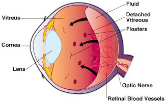
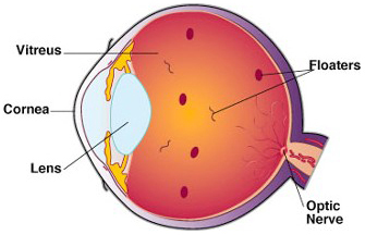
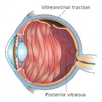
About Flashes and Floaters.What are Flashes?Flashes of light lasting a few seconds may appear in your vision when the vitreous gel pulls or tugs on the retina. This may happen as a natural result of aging or it may occur temporarily if you receive a blow to the head or eye. Usually these flashes, which are often described as lightning streaks, are noticed at night. Light flashes appearing as wavy lines in both eyes and lasting from a few minutes to half-an-hour, are usually a sign of an ocular migraine headache. Migraine-related flashes are often noticed in a lighted environment. Flashes of this nature are not a symptom of eye problems. The onset of new light flashes of short duration at night, especially when accompanied by the appearance of many new floaters or a blackening out of part of your field of vision, may indicate a retinal tear or detachment.
Retinal detachment requires immediate medical attention, as it can easily cause blindness. Both the appearance of flashes and the sudden onset of numerous small floaters warrant an ophthalmological investigation. What are Floaters?Floaters are deposits of various size, shape, and consistency floating within the normally transparent fluid (vitreous) inside the eye, appearing as spots, threads, or fragments of cobwebs singly or together with several others in one's field of vision — especially when looking at a blank surface or an open monochromal space, such as blue sky. 
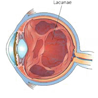
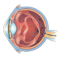
Despite the name "floaters", many of these specks have a tendency to sink toward the bottom of the eyeball, in whichever way the eyeball is oriented; looking up or lying back tends to concentrate them near the the center of one's gaze, while the textureless and evenly lit sky forms an ideal background against which to view them . Sometimes fine dark lines in amorphous mass - like small branching twigs - are seen. These floaters move around and are called 'muscae volitantes' (Latin for 'flying flies'), because they seem to dart about like flies as the eye is moved. Over time, you will become less aware of these floaters as the brain learns to ignore these retinal images. Therefore, while some floaters may remain in your vision, many of them will fade over time and become less bothersome. The Location of FloatersThe vitreous is a normally clear, gel-like substance that fills the center of the eye. It makes up approximately 2/3 of the eye's volume, giving it form and shape before birth.
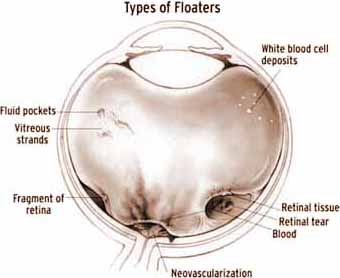
Floaters, when present, are suspended in the vitreous. Thus, they generally follow the rapid motions of the eye, while drifting slowly within the fluid. When they are first noticed, the natural reaction is to attempt to look directly at them. However, attempting to shift one's gaze toward them can be difficult since floaters follow the motion of the eye, remaining to the side of the direction of gaze. Floaters are, in fact, visible only because they do not remain perfectly fixed within the eye. Although the blood vessels of the eye also obstruct light, they are invisible under normal circumstances because they are fixed in location relative to the retina, and the brain "tunes out" stabilized images due to neural adaptation. This does not occur with floaters and they remain visible. The shapes are shadows projected onto the retina by tiny structures of protein or other cell debris discarded over the years and trapped in the vitreous humour. Floaters can even be seen when the eyes are closed on especially bright days, when sufficient light penetrates the eyelids to cast the shadows. The Occurrence of FloatersFloaters are common and do not cause problems for most people. In fact, people usually learn to ignore them, given some time. However, to those with severe cases, floaters are definitely a distraction — especially if the spots seem to constantly drift through the field of vision. Floaters have been known to catch and refract light in ways that somewhat blur vision temporarily until the floater moves to a different area. Many times they trick the sufferer into thinking they see something out of the corner of their eye that really is not there. For people with severe floaters, it is nearly impossible to completely ignore the large masses that constantly stay within almost direct view. Some sufferers have noted a decrease in ability to concentrate while reading, watching television, walking outdoors, and driving, especially when tired. Although more common in elderly people, floaters can certainly become a problem to younger people, especially if they are myopic. Floaters are also more common after cataract operations or after trauma. In some cases, floaters are congenital. Tear Film Debris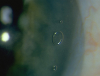
Sometimes the appearance of floaters has to be attributed to dark specks in the tear film of the eye. Technically, these are not floaters, but they do look the same from the viewpoint of the patient. People with blepharitis or a dysfunctional meibomian gland are especially prone to this cause, but ocular allergies or even the wearing of contact lenses can cause the problem. To differentiate between material in the vitreous humour of the eye and debris in the tear film, one can look at the effect of blinking: debris in the tear film will move quickly with a blink, while floaters are largely unresponsive to it. Tear film debris is diagnosed by eliminating the possibility of floaters and macular degeneration. Causes of FloatersThere are other causes for the appearance of floaters, of which the most common are described here. Basically, any way by which material enters the vitreous humour is a cause for floaters.
Diagnosis of FloatersUsually the appearance of new floaters or light flashes does not indicate any serious eye problem. However, the only way to ensure that the floaters or flashes are not symptomatic of a more serious problem, is to have your retina examined. If, following the exam, you develop large numbers of new floaters that seem to get worse over time, we recommend that you have your eyes re-examined. Standard vision tests like the Snellen visual acuity measurement, which measures your vision as 20/20, etc., are unable to quantify floaters and how the disability interferes with day-to-day functioning and overall quality of life. Floaters are often readily observed with the use of an ophthalmoscope or slit lamp. However, if the floater is a small piece of debris and near the retina, your eye doctor may not be able to observe it even if it appears large and obvious to you. Increasing background illumination or using a pinhole to effectively decrease pupil diameter may allow a person to obtain a better view of his or her own floaters. The head may be tilted in such a way that one of the floaters drifts towards the central axis of the eye. In the sharpened image the fibrous elements are more conspicuous. (If the pinhole is kept moving slowly in small circles, the same technique evokes an interesting entoptic effect known as the vascular figure, which is a view of the blood vessels within one's own eye.) Treatment of FloatersWhen floaters appear in your line of vision, move your eye around — up and down as well as from side to side. This movement creates a swirling in the vitreous fluid and may cause the floater to move out of your field of vision.
|
 (856) 667-3937Kindermann Eye Associates |
Eye Doctor South Jersey Kindermann Eye Associates - 3001 Chapel Avenue, Suite 200, Cherry Hill, New Jersey 08002Excellence in eye care for patients seeking a quality caring eye doctor in South Jersey. Dr. W Reed Kinderman is a premier ophthalmologist, New Jersey eye surgeon, specializing in cataract surgery, refractive surgery, laser guided cataract surgery, Tecnis multifocal lens implant, ReStor intraocular lens, Crystalens, Toric intraocular lens, glaucoma, strabismic eye muscle disorders, ophthalmology, and the full spectrum of eye care in New Jersey, Delaware Valley, Philadelphia, Mullica Hill, Ashland, Echelon, Thorofare, Riverton and surrounding areas. |